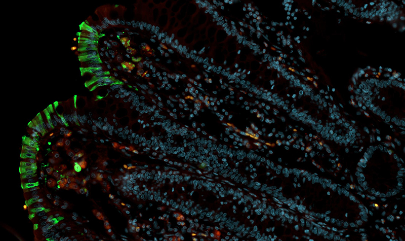
The Confocal Microscopy and Molecular Pathology Core is housed within the Division of Comparative Pathology.
The Confocal Microscopy and Molecular Pathology Core (CMMPC) provides research support and training with molecular pathology skills, confocal microscopy, image analysis, and multicolor immunohistochemistry and in-situ hybridization techniques for internal and affiliate scientists.
Services include assistance with experimental design, antibody selection, molecular probe selection, fixation and staining protocols, operating fluorescent and confocal microscopes, acquiring and storing imaging data, interpretation of results, and generation of publication quality images.
Image analysis services include assistance with capturing images for evaluation and with use of various image analysis software.
Contact
Cecily Midkiff, Lab Supervisor of the Confocal Microscopy and Molecular Pathology Core | cconerly@tulane.edu
Robert V. Blair, DVM, PhD, Diplomate ACVP | rblair3@tulane.edu
Acknowledgments
When utilizing research resources made possible by the TNPRC Confocal Microscopy Core, please cite RRID: SCR_024613. For additional information on acknowledging Tulane National Primate Research Center resources, please see here.
